Basic HTML Version


35
can easily prove that glucose in water is in the
4
C
1
conformation (as shown in
the figure).
1.2.10. Alginates: The
1
H-‐NMR
spectrum
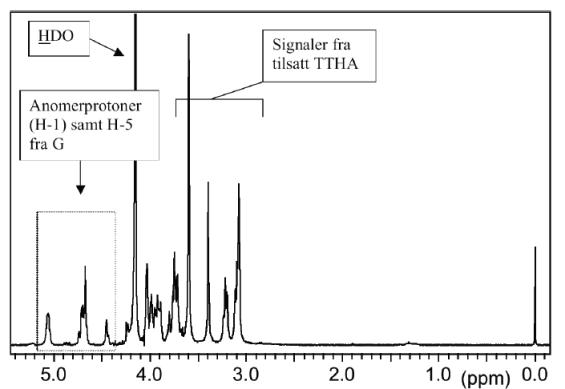
Figure 13
The
1
H-NMR spectrum of alginate was solved in a
seminal article by professor Hans Grasdalen
(Trondheim, Norway)
10
. The study was based on
several carefully prepared and characterized (by
chemical methods) alginate fragments with different
compositions and block structures. A typical alginate
spectrum is shown above.
Note that TTHA is added to bind traces of Ca
++
,
which would otherwise bind to the alginate and
influence the measurements. Before analysis the
alginate is degraded to DP = 50 (ca.). The spectra are further recorded at
90
°
C. In both cases this is done in order to obtain faster molecular motions
and hence, sharper peaks.
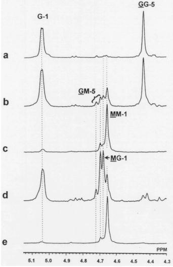
The figure (right) shows part (4.3-5.1 ppm region) of the
1
H-NMR spectra of
fragments of different alginates.
a)
Essentially purified G-block
b)
High G alginate (
L. hyperborea
)
c)
High M alginate (
P. aeruginosa
)
d)
Purified MG block (
A. nodosum
)
e)
Purified M block (
A. nodosum
)
10
H. Grasdalen (1983) High-field,
1
H-n.m.r. spectroscopy of alginate: sequential
structure and linkage conformations. Carbohydr. Res.
118
, 255-260.
Figure 14.
1
-H NMR spectra of
the anomeric region of four
alginates. a: G-block, b: high G
alginate, c: ca 50% M alginate,
e: M-block