Automating heart monitoring
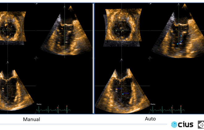
Heart disease is a major public health issue, and every year, more than 100,000 heart valve operations are performed in the United States alone. With these kinds of numbers, it is more and more urgent to simplify diagnosis and monitoring of heart patients. I/we believe automating the interpretation of ultrasound results is the way forward.
Assessing aortic stenosis severity by ultrasound
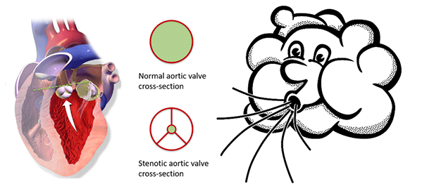
Aortic valve stenosis is a narrowing of the valve that separates the left ventricle from the aorta. A reduced opening increases the effort required by the left ventricle to pump blood. Being a degenerative disease, patients with aortic stenosis must undergo a clinical follow-up, which is usually performed by ultrasound. At the Centre of Innovative Ultrasound Solutions (CIUS), we are developing a new method that exploits 3-D high frame-rate imaging to increase the degree of automation in aortic stenosis flow measurements. The goal is to speed up the workflow in the clinics and increase the accuracy of measurements.
Improving ultrasound images of the heart’s blood vessels
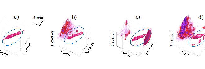
Coronary heart disease is a condition where the heart muscle (myocardium) does not receive enough oxygen and nutrients due to obstruction of blood flow in the heart’s blood vessels, known as the coronary arteries. It is therefore important to investigate the blood flow in these vessels. However, it is challenging to obtain accurate measurements due to the combination of poor blood flow, the constant movement of the heart throughout the cardiac cycle, and surrounding tissue causing noise signals.
Measuring the heart’s blood flow behaviour in 3D
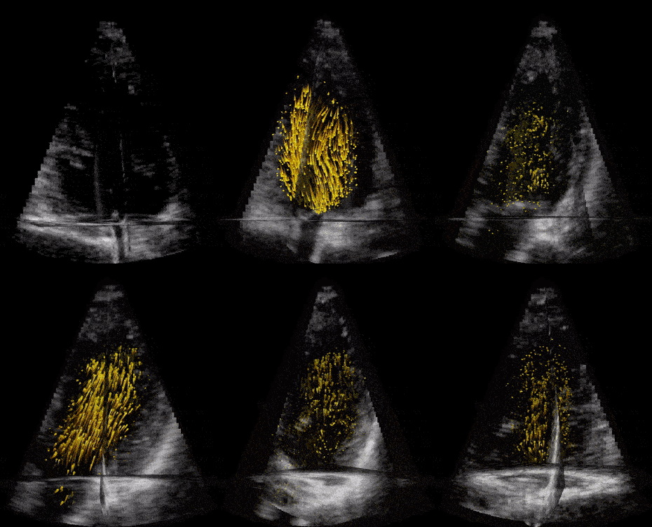
Given that cardiovascular related diseases are the most probable cause of death globally, according to WHO, we believe that more information regarding blood behaviour can help the doctors make better diagnosis at an earlier stage. But how can you measure these properties inside the heart, behind the ribs, under the skin, without moving the patients from their bed?
By Morten Smedsrud Wigen, PhD Candidate, Department of Circulation and Medical Imaging and CIUS – Centre for Innovative Ultrasound Solutions.
