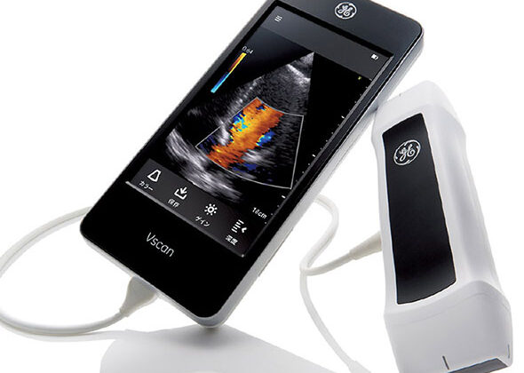
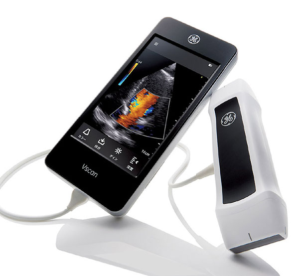
Could your local doctor diagnose heart disease using a handheld ultrasound device?
Many potential heart patients that are referred to specialists, turn out not to need specialist care. If general practitioners (GPs) could use handheld ultrasound devices with built-in diagnostic tools, could this improve patient outcome and reduce cost for the health services?
Improving ultrasound images of the heart’s blood vessels
Coronary heart disease is a condition where the heart muscle (myocardium) does not receive enough oxygen and nutrients due to obstruction of blood flow in the heart’s blood vessels, known as the coronary arteries. It is therefore important to investigate the blood flow in these vessels. However, it is challenging to obtain accurate measurements due to the combination of poor blood flow, the constant movement of the heart throughout the cardiac cycle, and surrounding tissue causing noise signals.
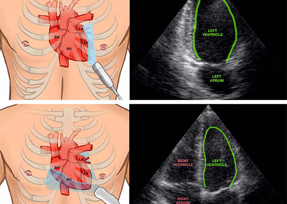
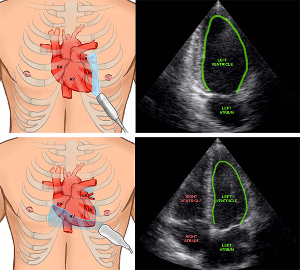
Using artificial intelligence to measure the heart
Artificial intelligence can now help clinicians by automatically measuring the heart in ultrasound images. This can save time and may in the future enable inexperienced users to perform accurate measurements of the heart.
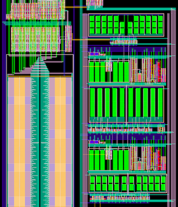
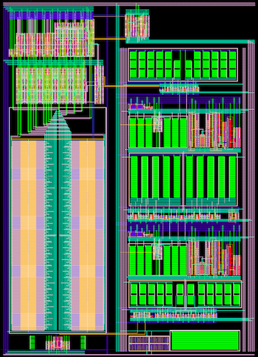
Making analog-to-digital converters for digital ultrasound probes
Making good digital ultrasound probes is extremely challenging because of the constrained amount of power that can be used. If significantly more than a couple of watts are consumed, the probe gets too hot to be allowed to touch your skin.
DeepEcho: Machine learning for improved echocardiography
Increased availability of ultrasound devices is great news, but it also triggers a range of new challenges. At CIUS we believe modern machine learning methods, such as deep learning, can provide necessary solutions.
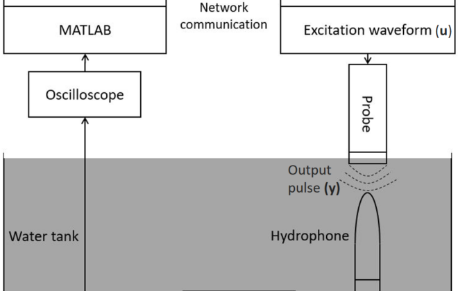
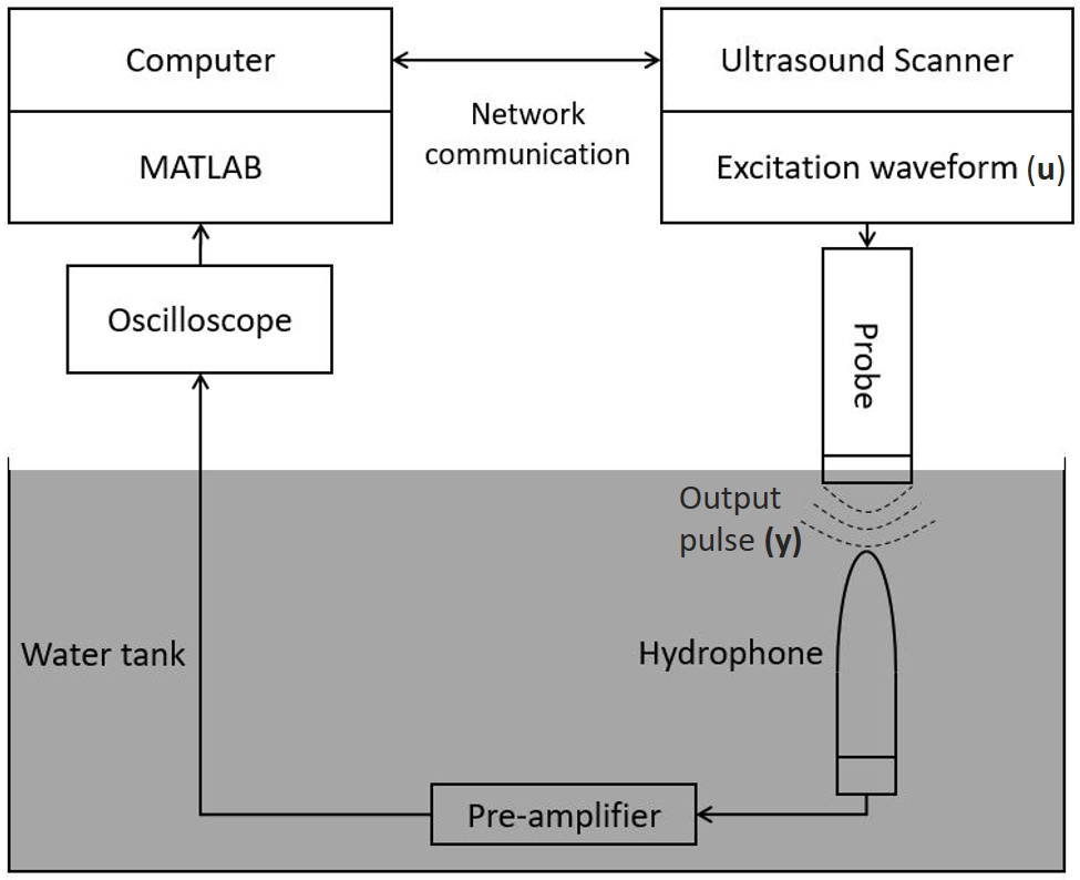
Optimization of transmit ultrasound pulses in second harmonic imaging
Today, second harmonic imaging is a preferred ultrasound imaging modality, mainly due to its ability to improve image quality by suppressing undesired acoustic reverberations. This imaging modality improves the diagnostics for different medical applications, and most of the cardiac ultrasound scanners in the hospitals use this method. We have established an experimental measurement system to measure, manipulate, and optimize excitation waveforms, and one of the goals is to reduce the undesired transmitted signal.
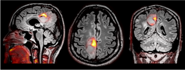
Can hybrid PET/MRI improve the diagnostic accuracy of brain glioma?
Gliomas are the most common primary brain tumors in adults with an incidence of around 250 cases per year in Norway. The outcome is poor, and the diagnostic accuracy is essential to estimate overall prognosis and to plan the best treatment for the patient. Can a hybrid PET/MRI system improve the diagnostic accuracy and treatment planning for this patient group?
Want to learn more about the Norwegian ultrasound research and business community?
Then watch NRK2 Saturday January 27th at 5.55 pm (in Norwegian) were I will talk about the development of the Norwegian ultrasound research and business community, and some of the ongoing projects within CIUS. The Norwegian ultrasound community is world leading within its fields. The…
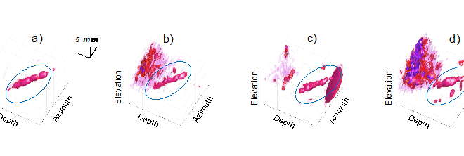
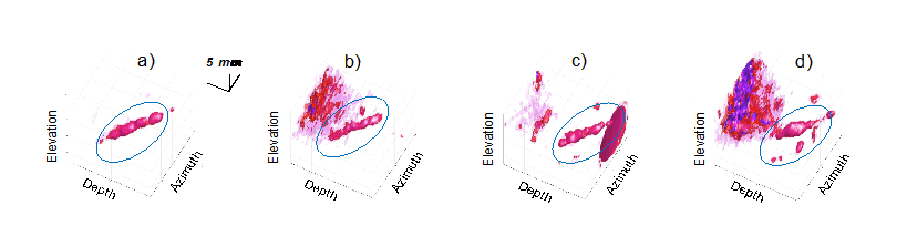
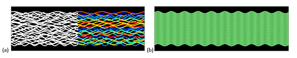
Measuring the heart’s blood flow behaviour in 3D
Given that cardiovascular related diseases are the most probable cause of death globally, according to WHO, we believe that more information regarding blood behaviour can help the doctors make better diagnosis at an earlier stage. But how can you measure these properties inside the heart, behind the ribs, under the skin, without moving the patients from their bed?
By Morten Smedsrud Wigen, PhD Candidate, Department of Circulation and Medical Imaging and CIUS – Centre for Innovative Ultrasound Solutions.
Enhancing marine sonar and medical ultrasound imagery using wave coherence
CIUS researchers are investigating whether a property of ultrasonic waves known as coherence can be used to detect microcalcifications in human tissue for cancer screening. In marine sonar, it has already been used successfully for detecting and characterising objects and features on the seafloor. The…