Improving quality of cardiac ultrasound images
Cardiovascular diseases (CVDs) are the number-one cause of death globally. An estimated 17.5 million people died from CVDs in 2012, 31% of all global deaths. One important factor in preventing cardiovascular disease, is early diagnosis using technologies such as ultrasound echocardiography. In this project, we study the cause of certain defects in the current echocardiograms and will try to propose new processing methods to improve the image qualities.
The quality of cardiac ultrasound images (echocardiograms) has increased significantly in the last 20 years, making it possible to correctly diagnose the occurrence of CVDs in about 80% of patients. In the remaining 20% certain physical factors hinder a correct visualization of the heart and the assessment of its function.
One of the artefacts which is seen in the echocardiograms taken from some patients, is what we refer to as “haze” artefact. In the images with this artefact a haze-like noise can be seen over the heart. This haze-like noise normally disappears when imaging deeper than 10 cm into the body in the studied cases (see the hazy echocardiogram in the figure below). In the following figure a “non-hazy” echocardiogram is shown together with a “hazy” one.

Echocardiography produces real-time images of the heart, by sending ultrasonic pulses (sound waves with higher frequency than audible frequency range) in between the ribs and down into the body. Ultrasonic pulses are then reflected back to the transducer (the transmitter and receiver of the ultrasonic pulses), which can be detected and transformed into an image. However that is not the only path that the signal can take. Sometimes the signal can be partially blocked by the ribs impacting the quality of the result images. Therefore, as a potential cause of the haze artefact, we investigate the geometry of rib bones and its effect on the received ultrasound signal from the heart.
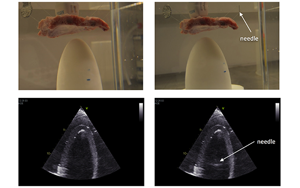
To study this effect, we carried out an experiment out of the body, where we imaged an artificial ventricle in a water tank. A section of a pork ribcage was placed on top of it to simulate the ribs effect in human body (see figure below). We imaged the ventricle through the ribs with an M5Sc transducer and E95 GE scanner. We repeated the imaging after placing the tip of a metal needle under water about 10 cm away from the transducer. Figure below shows the setup with and without the needle and the corresponding ultrasound images. We observe that the needle tip is displayed in the ultrasound image at a depth of around 11 cm (see the lower right pane in the figure below). However the needle is not expected to be visible in the image since it is placed out of the transducer field of view or “imaging plane” (a cross section of the object which is being imaged).

This experiment shows that if the ultrasound beam is partially blocked by the ribs, then part of the energy is reflected to unwanted directions. This deflected energy can then be reflected once more by the scatterers that are present in these directions (the needle tip in our experiment) and travels back to the transducer. Therefore, the scatterers out of the imaging plane can be observed in the ultrasound image as noise. The result of this experiment can be expanded to the haze artefact in the echocardiograms with the hypothesis being:
In some patients, with a specific shape and angle of the ribs, and depending on the heart position relative to the ribs, there is no way for all of the ultrasound beam to go through the ribs before hitting the desired cross section of the heart. This leads to the beam being partially reflected and any scattering tissue out of the imaging plane is then rendered as haze noise.
At this stage, we are collecting some live data from volunteers to check the haze level in their echocardiograms. At the same time, we record some data of the shape and distance of their ribs and compare the haze level with this information.

Ali Fatemi
PhD Candidate 2016-
- Ali Fatemi#molongui-disabled-link


